
Figure 3.
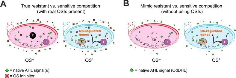
Comparison of a “true” QSI resistance competition to the experimental mimic competition in this study. “R” circles represent QS receptor proteins, and “I” circles represent QS signal synthase proteins. In the true case (A), the wild-type bacteria have chemically knocked down QS (red), and the resistant mutants are still capable of QS even in the presence of the QSI (blue). In the mimic case (B), a P. aeruginosa ΔlasR ΔrhlR mutant has a genetically knocked down QS system (red) to mimic the QSI-sensitive strain, and the resistant mimic is wild-type PAO1 (blue), which is fully capable of QS under the experimental conditions. In both panels, substantial native AHL signals are shown, but if the resistant bacteria are rare, much less signal will actually be present (due to poor signal production by the QS-inhibited strains).