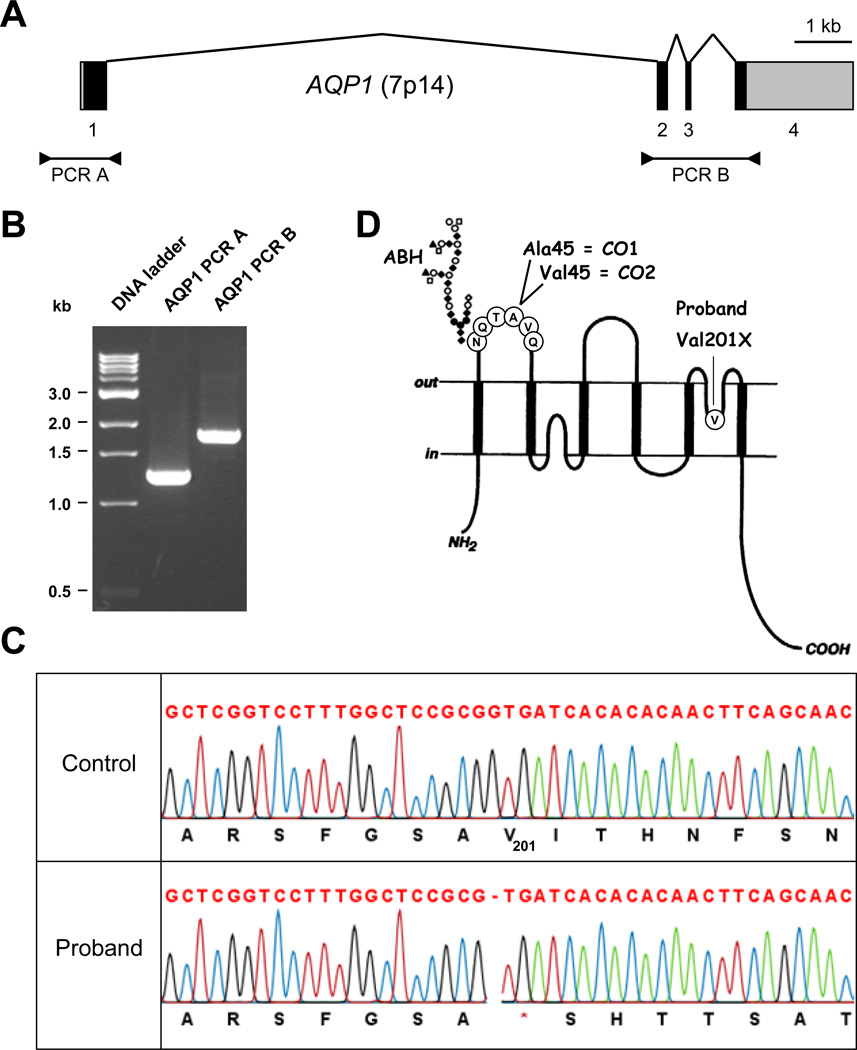
Figure 1. Identification of an AQP1 mutation in the proband.
(A) Diagram showing the structure of AQP1 and the two fragments that were PCR-amplified for sequencing; exons are depicted as boxes (coding sequences are in black and untranslated regions in gray) and introns as broken lines; PCR products are depicted below.
(B) PCR products of the two AQP1 fragments used for sequencing and analyzed using a 1.5% agarose gel; PCR A contains the AQP1 proximal promoter and exon 1, while PCR B contains the rest of the AQP1 coding sequence (exons 2 to 4).
(C) Detail of AQP1 sequencing from the proband (lower traces) and a control (upper traces) showing the apparently homozygous mutation c.601delG, p.Val201X in the proband.
(D) Diagram showing the membrane topology of AQP1 and the localization of ABH, CO1 and CO2 antigens (adapted from [8]).

