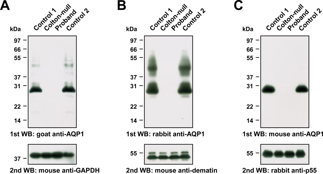
Figure 2. Immunoblot analysis of AQP1 from RBC membrane lysates prepared from the proband.
Equal amounts of membrane lysates prepared from the RBCs of two control donors (lanes 1 and 4), a previously characterized Conull subject (SAR, lane 2) and the proband (lane 3) were resolved by Tris-Glycine 12% SDS-PAGE electrophoresis. Samples were not reduced or heat-denatured prior to electrophoresis, and were transferred to PVDF membrane for immunoblot analysis.
(A) PVDF membrane probed with goat anti-AQP1 (upper panel) and re-probed with mouse anti-GAPDH (lower panel).
(B) PVDF membrane probed with rabbit anti-AQP1 (upper panel; the smear migrating between 37 and 55 kDa corresponds to glycosylated AQP1) and re-probed with mouse anti-dematin (lower panel; erythroid dematin is expressed as two isoforms produced by alternative splicing).
(C) PVDF membrane probed with mouse anti-AQP1 (upper panel) and re-probed with rabbit anti-p55 (lower panel).

