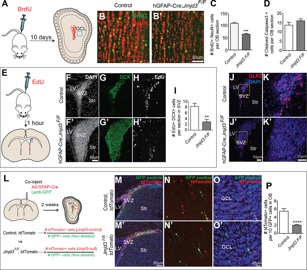
Figure 1. Jmjd3 is required for adult OB neurogenesis.
(A) Illustration of the experimental design for B-D. Green box indicates regions of shown in B and B’. GCL, granule cell layer. (B-D) Analysis of OB neurogenesis. (B and B’) Immunohistochemistry (IHC) for BrdU (green) and NeuN (red) in coronal OB sections of control (B) and hGFAP-Cre;Jmjd3F/F mice (B’). (C) Quantification of BrdU+, NeuN+ OB neurons (*** P<0.001, n=4 each, error bars, s.e.m). (D) Quantification of Caspase3+ OB cells (P=0.1933, n=4 each, error bars, s.e.m.)
(E) Illustration of the experimental design for F-I. EdU was injected 1h before analysis. (F-I) Analysis of cell proliferation and neuroblasts in the SVZ. (F-H) DAPI+ (white), DCX+ (green), and EdU+ (white) cells in the SVZ of control mice. (F’-H’) DAPI+ (white), DCX+ (green), and EdU+ (white) cells in the SVZ of hGFAP-Cre;Jmjd3F/F mice. (I) Quantification of EdU+, DCX+ cells in SVZ coronal sections (n=3 each, error bars, s.e.m., **P<0.01).
(J-K’) Analysis of DLX2 expression in the SVZ. IHC for DLX2 (red) in SVZ coronal sections of control (J-K) and hGFAP-Cre;Jmjd3F/F mice (J’-K’). Panels K and K’ are higher magnification views of the yellow boxed region in J and J’.
(L) Schematic illustration of the experimental design for M-P. Ad:GFAP-Cre virus (to delete floxed alleles in GFAP+ SVZ NSCs) and GFP lentivirus (injection control) were co-injected into the adult SVZ of tdTomato;Jmjd3F+ (M-O) or tdTomato;Jmjd3FF (M’-O’) mice. (M-P) Analysis of OB neurogenesis. (M and M’) IHC for GFP (green) and tdTomato (red) in adult SVZ coronal sections of tdTomato;Jmjd3F+ (M) and tdTomato;Jmjd3FF mice (M’). (N-O’) IHC for GFP (green) and tdTomato (red) in coronal OB sections of tdTomato;Jmjd3F+ (N and O) and tdTomato;Jmjd3FF mice (N’ and O’) 14 d after injection. (P) Quantification of tdTomato+ neurons per 10 GFP+ neurons in OB (**** P<0.0001; n=4 per group; error bars, s.e.m.). LV, lateral ventricle; Str, Striatum; GCL, granule cell layer.

