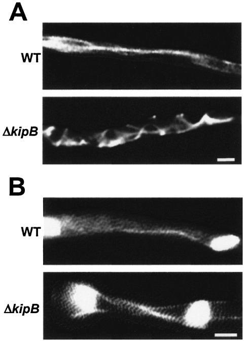
FIG. 4.
Morphology and MT organization in ΔkipB mutant strain and wild type. MTs were observed as α-tubulin-GFP fusions by fluorescence microscopy (see Materials and Methods). Top rows, wild type; bottom rows, ΔkipB strain. (A) Cytoplasmic MTs. In the wild type, they are long and straight, while they display a curved pattern in the ΔkipB mutant. (B) Mitotic spindles. In the wild-type spindle, the cytoplasmic MT remained, while in the ΔkipB strain, three filaments are visible. Bar, 5 μm.

