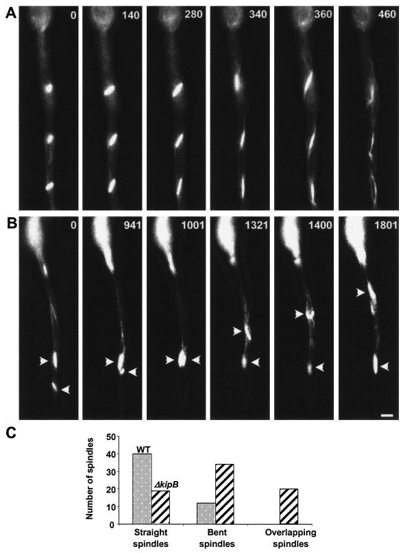
FIG. 5.
Positioning of mitotic spindles in the wild type and the ΔkipB disruptant. Images from a time-lapse series are displayed (times are indicated, in seconds, in the upper right corner of each panel). (A) Wild-type synchronized mitoses, with evenly distributed spindles along the length of the hypha. (B) Mitoses in the ΔkipB mutant strain, with defects in spindle positioning and morphology (sharp angled bow-like structures) and with a high level of spindle mobility through the cytoplasm, which leads to overlapping of the spindles (arrows). Bar, 5 μm. (C) Quantification of different spindle morphologies and spindle behavior in the wild type and the ΔkipB mutant strain (also see Videos S1 and S2 in the supplemental material).

