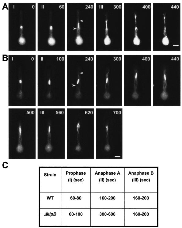
FIG. 6.
kipB disruption causes a delay in mitotic progression. The images show time-lapse analyses of mitosis in germlings of the wild type (strain GFP-tubA) (see Video S3 in the supplemental material) (A) and the ΔkipB mutant (SPR30) (see Video S4 in the supplemental material) (B). MTs were labeled with GFP. The stages of mitosis are indicated in the upper left corner of the pictures. I, prophase to metaphase (short spindle); II, anaphase A (spindle elongates very slowly, with the appearance of astral MTs [indicated by arrows]); III, anaphase B (spindle elongates rapidly and doubles or triples in length). The cells were grown overnight at 30°C and were observed at room temperature. Images were taken every 20 s, and a selection of them are displayed here. The time points (in seconds) are indicated in the upper right corner of the pictures. Bar, 5 μm. (C) Summary of the time intervals of different mitotic phases in the wild-type and kipB mutant strains (see Videos S3 [wild type] and S4 [mutant] in the supplemental material).

