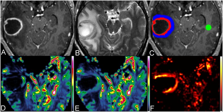
Figure 1. Measurements of perfusion parameters in a 47-year-old man with pyogenic brain abscess.
Axial contrast-enhanced MPRAGE (A) and T2W image (B) show a rim-enhancing mass with perifocal edema in the right temporal lobe. (C) On contrast-enhanced MPRAGE, three ROIs are placed over the enhancing rim (red), perifocal edema (blue) most adjacent to the enhancing rim and the contralateral NAWM (green) for the measurements of CBV (D), corrected CBV (E) and K2 (F), respectively.

