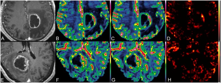
Figure 2. MR perfusion of a glioblastoma and a metastatic brain tumor.

The upper panel shows the contrast-enhanced MPRAGE (A), CBV (B), corrected CBV (C) and K2 (D) images of a necrotic glioblastoma in the left medial parietal region, and the lower panel (E, F, G and H) shows the corresponding images of a cystic metastatic brain tumor in the right occipital lobe.
