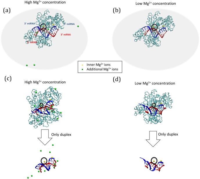
Figure 1. Three-dimensional structures of the miRNA-mRNA-Ago2 complex.
The initial Ago2 structures under high and low concentrations of Mg2+ ions (Ago-high and Ago-low) are presented in (a) and (b), respectively. The final structures of the complex and the duplex after 10 ns of simulation are shown for high (c) and low (d) concentrations of Mg2+ ions. Two replicates (HM4 for Ago-high and LM2 for Ago-low complex) are used for drawing. The components of the complex are the Ago2 protein (cyan tube), mRNA (red strand), the miRNA (target RNA) (blue strand), inner Mg2+ ions (yellow spheres), and outer Mg2+ ions (green spheres, only in Ago-high). The black circle represents the Mg2+ ion in the catalytic region interacting with the target RNA (red strand). In the miRNA-mRNA duplex, the bases are drawn as tube models. The periodic box for the computational simulation is not shown.

