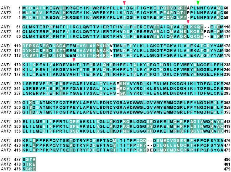
Figure 3. Multiple Sequence Alignment of the three AKT isoforms, AKT1, AKT2, and AKT3.

The conserved positions are shown in light green and the corresponding amino acids in black font, whereas the less-conserved positions are shown in gray color with the corresponding amino acids in white font. The initial and final position of each isoform in all the rows of the alignment is also provided. The position-equivalent-residues (residues of different isoforms falling at same column position in the isoform alignment) overlapping among the interacting residues of MK-2206 are marked by triangles; the green triangle (Asn-53) indicates the residue overlapping between AKT1 and AKT2 binding, while the red triangles indicate the position-equivalent-residues overlapping among the interacting residues of AKT2 and AKT3 which are Ser-31, Leu-110, and His-196 of AKT2 (corresponding to Thr-31, Leu-109, and His-192 of AKT3 respectively).
