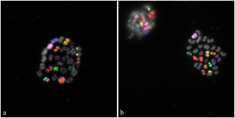
Figure 4. M-FISH carried out on bovine in vitro maturated secondary oocytes.
Specific fluorescent signals were identified on: a) oocyte at the diakinesis/metaphase I stage of the meiosis; b) oocyte at MII and corresponding PB I. Correct chromosomal segregation can be clearly indicated for 11 autosomes and X chromosome.

