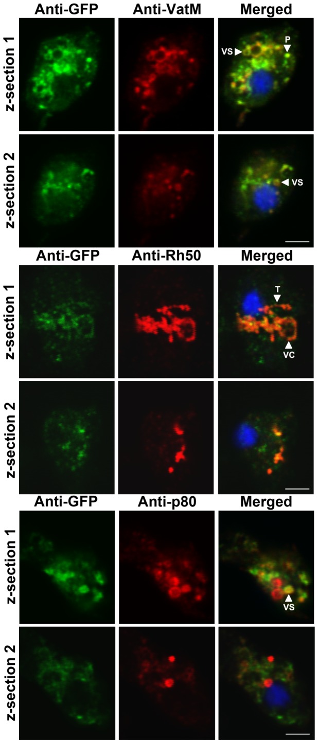
Figure 3. Intracellular localization of Dictyostelium GFP-Cln3 using confocal microscopy.
AX3 cells overexpressing GFP-Cln3 were fixed in either ultra-cold methanol (for VatM and Rh50 immunostaining) or 4% paraformaldehyde (for p80 immunostaining) and then probed with anti-GFP (rabbit polyclonal anti-GFP for anti-VatM and anti-p80 co-staining and mouse monoclonal anti-GFP for anti-Rh50 co-staining) followed by anti-rabbit or anti-mouse Alexa 488. Cells were then probed with one of anti-VatM, anti-Rh50, or anti-p80 followed by the appropriate secondary antibody linked to Alexa 555. Two z-sections are shown for each cell. Z-sections 1 and 2 are approximately 1 µm and 3 µm, respectively, from the bottom of each cell. VC, vacuolar-shaped structures; VS, cytoplasmic vesicles; T, tubular-like structures within the cytoplasm; P, punctate distributions within the cytoplasm. Scale bars = 2.5 µm.

