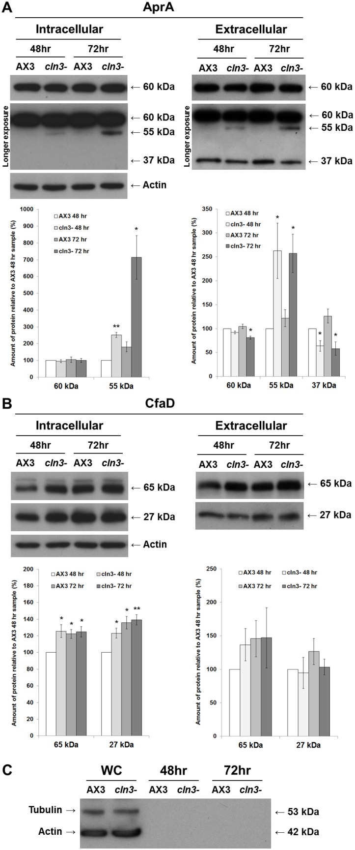
Figure 6. Effect of cln3 knockout on the intra- and extracellular levels of AprA and CfaD.
AX3 and cln3− cells grown axenically in HL5 were harvested and lysed after 48 and 72 hours of growth. Whole cell lysates (20 µg) (i.e., intracellular) and samples of conditioned growth media (i.e., extracellular) were separated by SDS-PAGE and analyzed by western blotting with anti-AprA, anti-CfaD, anti-tubulin, and anti-actin. Molecular weight markers (in kDa) are shown to the right of each blot. (A) Intra- and extracellular protein levels of AprA. Immunoblots that were exposed for a longer period of time (i.e., longer exposure) are included to show the 55-kDa and 37-kDa protein bands detected by anti-AprA. Note that the 37-kDa protein was detected in samples of conditioned growth media, but not in whole cell lysates. (B) Intra- and extracellular protein levels of CfaD. Data in all plots presented as mean amount of protein relative to AX3 48 hour sample (%) ± s.e.m (n = 4 independent experimental means, from 2 replicates in each experiment). Statistical significance was determined using a one-sample t-test (mean, 100; two-tailed) vs. the AX3 48 hour sample. *p-value<0.05. **p-value<0.01. (C) Detection of tubulin and actin in whole cell lysates (WC; lanes 1–2), but not in samples of conditioned growth media (lanes 3–6).

