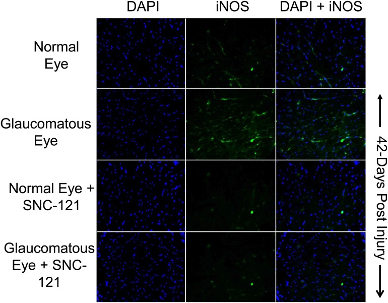
Figure 8. Changes in iNOS expression in glaucomatous optic nerve at 42 days, post injury.
The optic nerve (non-myelinated; 2 mm post globe) of Brown Norway rats was removed 42-days post glaucomatous injury. Contralateral optic nerve was used as the normal control. Cryosections were immunostained by anti-iNOS antibodies as indicated horizontally. Ocular treatments are indicated vertically. Data shown in this Figure are a representation of at least four independent experiments. A total of 10 animals were used in this experiment. Comparable staining for iNOS was seen in at least 4 animals in each treatment group.

