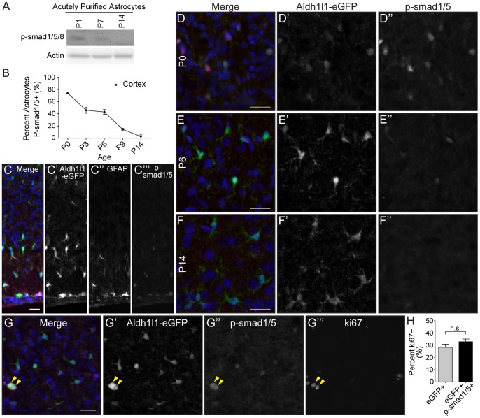
Figure 4. Cortical astrocytes in vivo display smad1/5/8 pathway activation during early postnatal development.
A. Western blotting for p-smad1/5/8 in acutely purified astrocytes across developmental time points demonstrates pathway activation during the early postnatal period. Actin bands confirm equal protein loading. B–F. Immunostaining of sagittal brain sections from Aldh1l1-eGFP mice to visualize astrocytes. Merged images show DAPI (blue), p-smad1/5 (red) and Aldh1l1-eGFP (green) (D–F). Colocalization of p-smad1/5 with eGFP+ cortical astrocytes was quantified across the first two weeks of postnatal development using ImageJ (B). N = 2 animals for each time point. Immunostaining of P6 brains sections with DAPI (blue), p-smad1/5 (red), GFAP (magenta) and Aldh1l1-eGFP (green) shows that p-smad1/5 and GFAP staining localizes predominately to astrocytes in the outer cortical layers and glia limitans (C). G–H. Sagittal brain sections from Aldh1l1-eGFP mice were sequentially immunostained for p-smad1/5 and ki67. Yellow arrows indicate cortical astrocytes that express both p-smad1/5 and ki67 (G). Colocalization of p-smad1/5 and ki67 with eGFP+ cortical astrocytes was quantified using ImageJ (H). N = 3 animals. Significance determined using students t-test. All scale bars are 25 µm. Error bars represent SEM.

