
Figure 4. Effect of EGTA and ionomycin on the expression of pro- and anti-inflammatory cytokines in L. donovani infected macrophages.
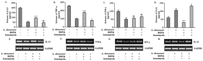
Macrophages (2×10⁶ cells) were infected with L. donovani (1∶10) and incubated at 37°C in the presence of 2 mM EGTA and/or 1 µM ionomycin. The cell supernatants were collected after 24 hr of infection. Cytokine assay was carried out by ELISA (A, B, C and D). Data represent means ± SD for 3 sets of experiments ***P<.001, *P<.05 and ns = non-significant for the comparison with infected one. In a parallel experiment, EGTA or Ionomycin treated infected cells were collected in Trizol for RNA extraction and semi-quantitative RT-PCR was performed. IL-12, TNF-α, IFN-γ, IL-10 and GAPDH PCR products were resolved on an agarose gel (1.5%) and quantified densitometrically using lab software as described in Methods. Data is from one representative experiment performed at least three times (E, F, G, and H).