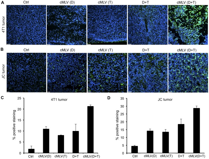
Figure 5. Effect of codelivered cMLVs on tumor apoptosis (D: Dox; T: PTX).
(A, B) Mice bearing either 4T1 tumor (A) or multidrug-resistant JC tumor (B) were injected intravenously through the tail vein with cMLV (5 mg/kg Dox), cMLV (5 mg/kg PTX), 5 mg/kg Dox + 5 mg/kg PTX, or cMLV (5 mg/kg Dox+5 mg/kg PTX). Three days after injection, tumors were excised. Apoptotic cells were detected by a TUNEL assay (green), followed by nuclear costaining with DAPI (blue). Scale bar represents 50 µm. (C, D) Quantification of apoptotic cells in 4T1 (C) and JC (D) tumors. To quantify TUNEL-positive cells, 4 regions of interest (ROI) were randomly chosen per image at ×2 magnification. Within one region, area of TUNEL-positive nuclei and area of nuclear staining were counted. The data are expressed as % total nuclear area stained by TUNEL in the region. Data are represented as mean ± SD (n = 3).

