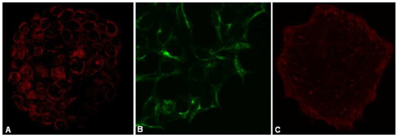
Figure 1. Phenotyping cultured human cardiomyocytes.
Human cardiomyocytes cultured on microscope cover slips where phenotyped using cardiac markers (A) α-MHC (63× magnification) (AF546-labeled secondary antibody [red]) (B) Cx43 (x40) (AF488-labeled primary antibody [green]) and (C) cTnT (40× magnification) (AF546-labeled secondary antibody [red]). Cardiomyocytes where imaged on a Zeiss 780 immunofluorescent confocal microscope.

