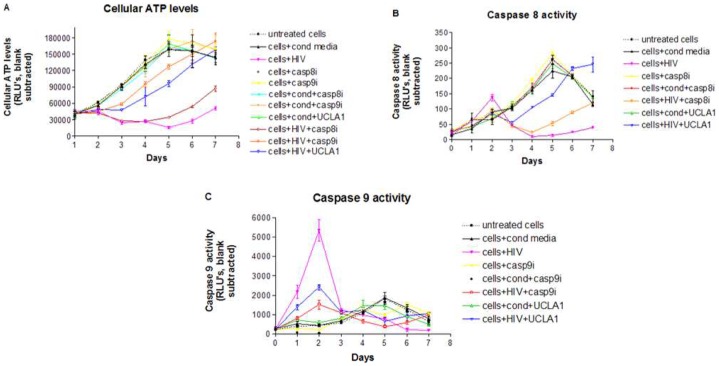
Figure 5. Caspase 8 and 9 activities in CM infected with HIV-1.
CM were exposed to: media alone; conditioned media containing virus; HIV; 100 µM of casp8i; 100 µM of casp9i; conditioned media and 100 µM of casp8i; conditioned media and 100 µM of casp9i; conditioned media and 100 nM UCLA1 aptamer; HIV and 100 µM of casp8i; HIV and 100 µM of casp9i; HIV pre-incubated for 1 h with 100 nM of UCLA1 aptamer. Cells were harvested daily for 7 days. (A) ATP levels were measured as a correlate of cell viability; (B) caspase 8 and (C) caspase 9 activities were measured as indicators of extrinsic and intrinsic apoptosis, respectively. All assays were luminescence-based and presented as graphs relating RLU to number of days (n = 3± SEM).

