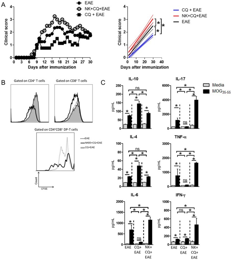
Figure 1. Aggravation of EAE in mice cured from malaria correlates with increased cellular immune response towards myelin.
C57BL/6 mice (n = 6 mice/group) were intraperitoneally (i.p.) infected with 1×10⁶ P.berghei-infected Red Blood Cells and treated with chloroquine (CQ, 5 mg/Kg) for five consecutive days starting at the 10th day after infection. Three days after the last dose of CQ, mice were immunized with 100 µg of MOG35–55 peptide and Pertussis toxin was administrated (via i.p.) at 0 and 48h after peptide immunization for EAE induction. A) The clinical course of EAE was then monitored. Linear regression analyses are exposed in the side panels, thinner lines indicate 95% confidence interval. B) At the 10th day after MOG-immunization, the spleens of mice were collected and dissociated. Total leukocytes (5×10⁵/well) were CFSE-stained (2,5 µM) and cultured in the presence of MOG35–55 (10µg/mL) peptide for 96h. At the end of culture period, the cells were surface stained with anti-CD3/CD4/CD8 antibody cocktail and events were acquired in a flow cytometer. The proliferation was analyzed inside each T cell population. C) The culture supernatants were assayed for the secreted cytokines IL-10, IL-4, IL-6, IL-17, TNF-α and IFN-γ. Data was analyzed by One-Way Anova and post-tested with Bonferroni. In all analyses, *: p<0,05; ns: not significant. Representative data of three independent experiments.

