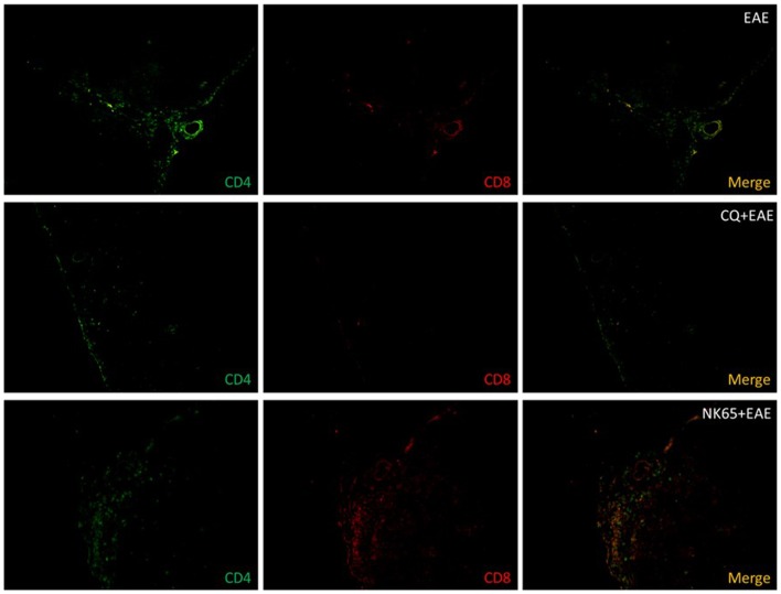
Figure 3. Central Nervous System of malaria-cured EAE mice show increased cellular infiltration of DP-T cells.
C57BL/6 mice (n = 6 mice/group) were intraperitoneally (i.p.) infected with 1×10⁶ P.berghei-infected Red Blood Cells and treated with chloroquine (CQ, 5 mg/Kg) for five consecutive days starting at the 10th day after infection. Three days after the last dose of CQ, EAE was induced. As controls, naïve mice were treated with CQ or vehicle before EAE induction. The spinal cords of EAE-inflicted mice were collected fourteen days after MOG-immunization. Frozen thin sections (12 µm) were made and fixed in formalin. Cells were stained with FITC-conjugated anti-CD4 and PE-conjugated anti-CD8 and analyzed in epifluorescence microscope. Figures are representative of three independent experiments. Magnification: 200X.

