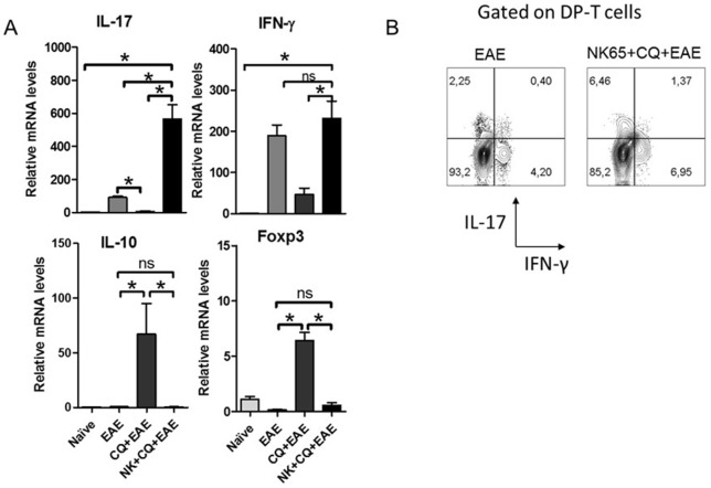
Figure 4. Inflammation in the CNS of NK+CQ+EAE mice correlates with an increased production of inflammatory cytokines by DP-T cells.
Groups of mice (n = 6 mice/group) subjected to infection and EAE induction. A) At the 10th day after MOG-immunization, mice were killed and spinal cords were removed to analyze the gene expression of IL-17, IFN-γ, Foxp3 and IL-10 in the lumbar spinal cords of mice. Data was analyzed by One-Way Anova and post-tested with Bonferroni. B) The infiltrating cells of the CNS were enriched and stimulated by Phorbol Myristate Acetate and Ionomycin in the presence of Brefeldin A for 4 h. The frequency of IFN-γ- and IL-17-producing cells inside CD4+CD8+ T cell gate was analyzed. In all analyses, *: p<0,05. ns: not significant. Representative data of three independent experiments.

