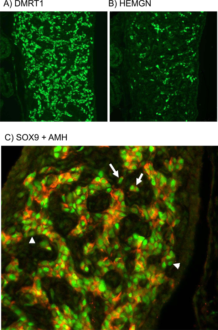
Fig. 2.
Localisation of key testis pathway proteins in developing male gonads. Immunostaining of E7.5 (HH32) male gonads. (A) DMRT1 (green) staining shows widespread expression in nuclei of Sertoli cells. (B) Serial adjacent section showing HEMGN (green) staining. Expression is also localised to Sertoli cell nuclei, but is of variably intensity and limited to a subset of cells. (C) Double staining of SOX9 (green) and AMH (red) in developing Sertoli cells, showing that these proteins are co-expressed in the vast majority of cells (e.g, white arrows). However some cells are SOX9 positive but AMH negative (e.g., white arrowheads).

