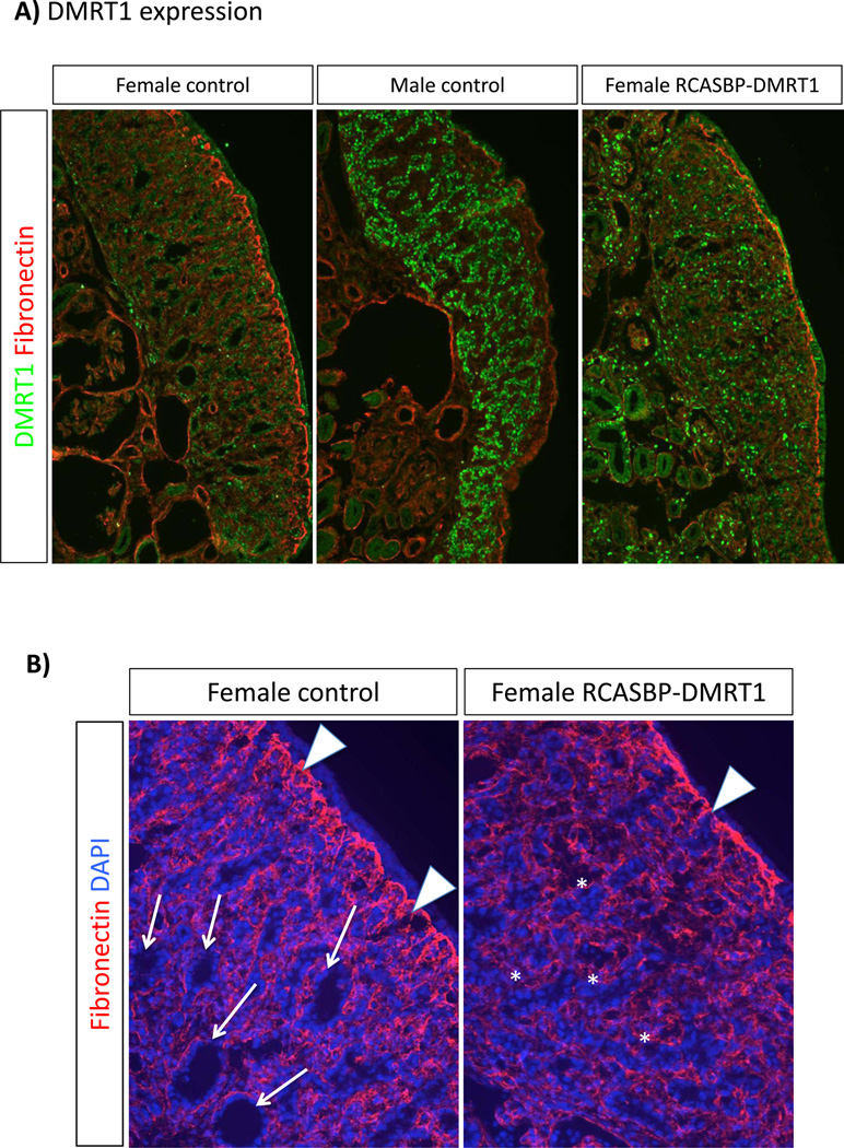
Fig. 4.
Structural effect of DMRT1 over-expression on gonadal development. Immunostaining on E7.5 left control gonads and left female gonads electroporated with RCASBP-DMRT1. All images were captures at the same exposure. (A) DMRT1 (green) and fibronectin (red) staining. Control females (not electroporated) show a low level of DMRT1 expression throughout the gonad, and a thickened fibronectin positive basement membrane between the outer cortex and underlying medulla. In male controls, DMRT1 expression is higher and localised to developing seminiferous cords, while the fibronectin layer is weaker. DMRT1 is over-expressed in female gonads infected with RCASBP-DMRT1, and the fibronectin layer is less well-developed than in the control female. (B) Fibronectin (red) and DAPI (blue) staining in control versus DMRT1- over-expressing female left gonads. In the control, numerous lacunae are evident in the medulla (e.g., arrows), while the fibronectin+ basement memebrane is extensive (white arrowheads) and the outer cortex beyond the BM is clear. In the female gonad over-expressing DMRT1, medullary lacunae are far less frequent, but medullary cells are arranged instead in a seminiferous cord like pattern (asterisks). Fibronectin+ basement membrane is less well developed (arrowhead) and the cortex itself is noticeably thinner.

