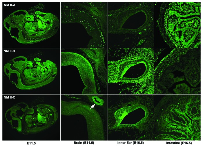
Figure 1. Expression of NM II in Mouse Embryos. Sections of paraformaldehyde-fixed mouse embryos were stained with antibodies to NMHC II-A (upper row), II-B (middle row), and II-C (lowest row). All three NM IIs are widely distributed throughout the E11.5 embryo (left column). In E11.5 mouse brain (second column), NM II-A is enriched in vasculature, NM II-B is the major isoform detected in neurons, and NM II-C stains intensively in developing pituitary (white arrow). In developing mouse inner ear at E16.5 (third column), NM II-A mostly stains the vasculature, II-B is detected in both mesenchymal and epithelial cells, and II-C is particularly enriched in the developing sensory cells of the cochlea. In the E16.5 mouse intestines (fourth column), both II-A and II-C are intensely stained in the epithelial cells, but II-C is particularly concentrated at the apical border of these cells; II-B appears more intense in the surrounding serosal cells.2

An official website of the United States government
Here's how you know
Official websites use .gov
A
.gov website belongs to an official
government organization in the United States.
Secure .gov websites use HTTPS
A lock (
) or https:// means you've safely
connected to the .gov website. Share sensitive
information only on official, secure websites.
