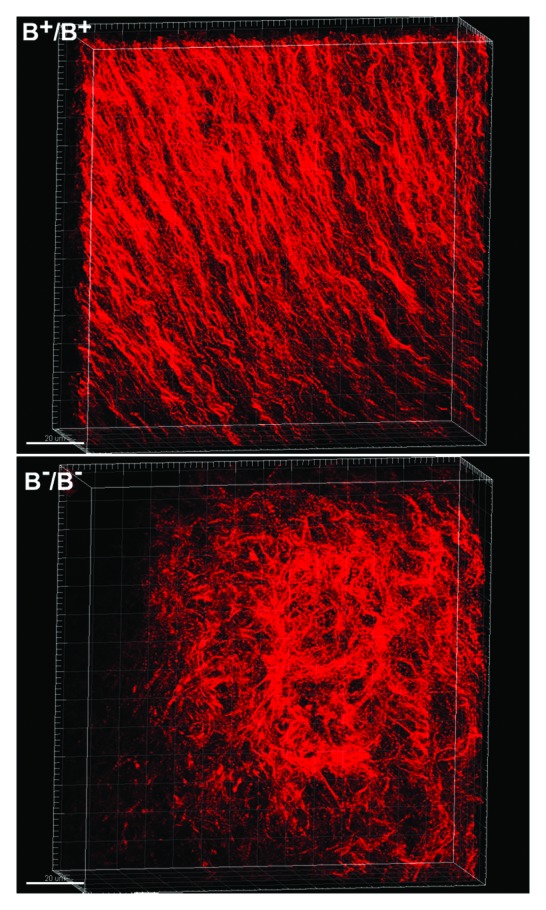
Figure 5. Disrupted Cardiomyocyte Alignment in NM II-B Ablated Mouse Hearts. E13.5 wild type (B+/B+) and NM II-B knockout (B-/B-) mouse hearts stained (wholemount) for actin-filaments with Alexa Fluor® 594 phalloidin. 3D stack of confocal microscope images of the ventricular myocardium shows that B+/B+ cardiomyocytes align in a regular spiral pattern (upper panel), while B-/B- cardiomyocytes are distributed randomly with no obvious pattern (lower panel).
