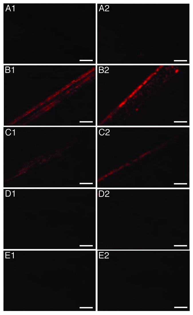
Figure 4. Fluorescence Microscopy of stainless steel wires. (A) uncontaminated, (B–E) incubated with 10% hamster brain homogenate infected with the 263K prion strain and treated with 224 μg mL−1 Fe3+, 500 μg mL−1 h−1 H2O2, pH = 3.5 and UV-A for 0, 240, 360, and 480 min respectively. Each row of panels (1–2) demonstrates areas of wires of the same group. Bar: 200 μm

An official website of the United States government
Here's how you know
Official websites use .gov
A
.gov website belongs to an official
government organization in the United States.
Secure .gov websites use HTTPS
A lock (
) or https:// means you've safely
connected to the .gov website. Share sensitive
information only on official, secure websites.
