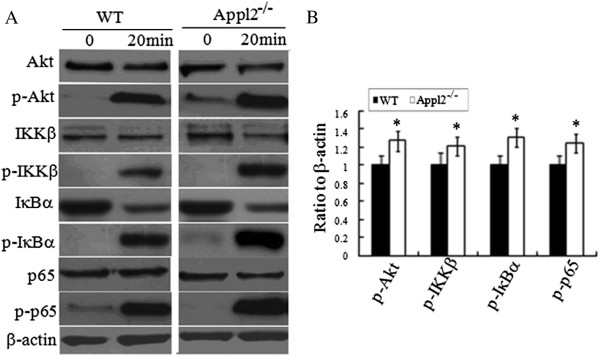
Figure 5.

Enhanced activation of Akt-NF-κB pathways after LPS stimulation in Appl2 KO macrophages. Peritoneal macrophages derived from peritoneal cells were stimulated with 1 μg/mL LPS for 20 min. The cell lysates were analyzed by western blot. (A) Western blot analysis on peritoneal macrophages from APPL2 KO mice with the indicated antibodies. (B) The ratio values of p-Akt/Akt/β-actin, p-IKKβ/IKKβ/β-actin, p-IκB/ IκB/β-actin, p-p65/p65/β-actin as determined by the density analysis of (A) using NIH Image J software for relative values. *p < 0.05, compared with WT mice, n = 5.
