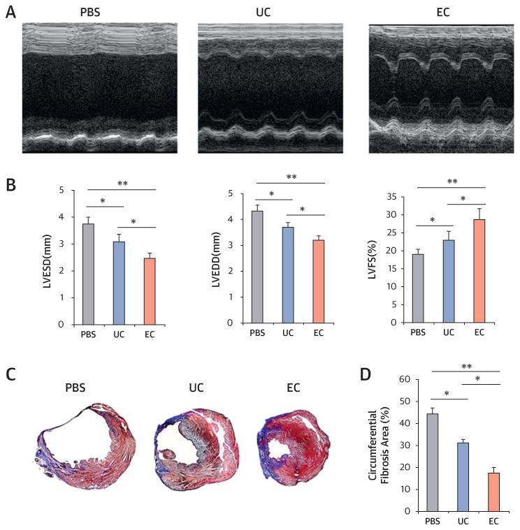
Figure 4. Endothelial Cell Transplantation Is Effective for Infarct Repair.

(A) Representative M-mode echocardiograms. (B) Left ventricular end-diastolic dimension (LVEDD) and left ventricular end-systolic dimension (LVESD) were lowest in the endothelial cell (EC) group. Left ventricular fractional shortening (LVFS) was better in EC-injected hearts compared with uncultured cell (UC)-injected or phosphate-buffered saline (PBS)-injected hearts at 3 weeks after myocardial infarction (n = 7 each; *p < 0.05). (C) Representative cross-sectional images of hearts stained with Masson’s trichrome at 4 weeks after cell transplantation. The blue color represents fibrosis. (D) EC transplantation offered the highest reduction in fibrosis area (n = 7; *p < 0.05; **p < 0.01).
