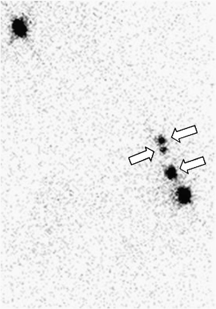
Fig. 3.
An image (anterior projection) acquired 22.3 h after a 1.2-mCi injection of unfiltered [Tc99m]sulfur colloid (Subject 8). Three sentinel lymph nodes (arrows) were detected. Four SLNs were excised 25.6 h postadministration. The primary SLN (lowest arrow) accumulated 7.52% of the dose and was positive for metastatic disease. The activity at the upper left corner is the imaging standard.

