
Fig. 3.
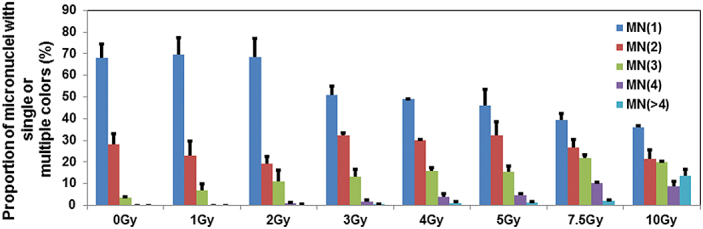
Percentages of micronuclei with single, double and multiple colours detected by M-FISH in cytochalasin B-blocked binucleated cells in mock and irradiated samples. The pooled data from the three donors are presented in the form of histograms. Bars indicate SEM.