Fig. 6.
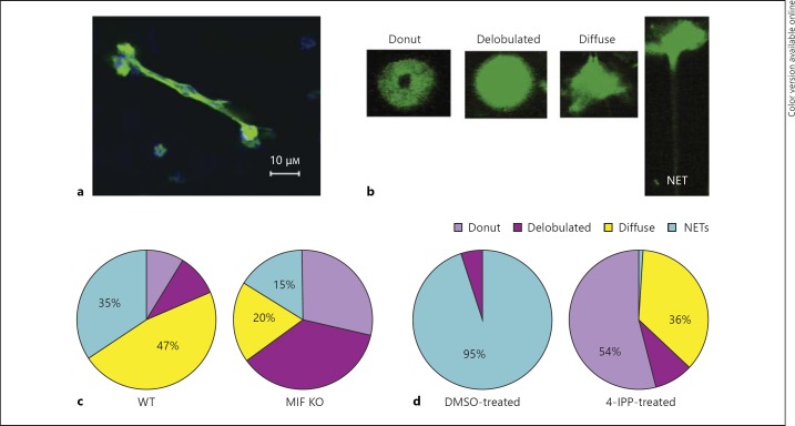
MIF promotes NET release. a NETs formed by C57BL6-derived PMNs in response to P. aeruginosa PAO1 stimulation. NETs were visualized by fluorescent DNA-specific staining with SYTOX Green in stimulated neutrophils after fixing and permeabilizing. The images were acquired on a Nikon confocal microscope with ×63 and ×40 objectives. b Stages of nuclear DNA rearrangement in response to P. aeruginosa stimulation of PMNs. The ‘donut’-shaped nuclei are typical of resting PMNs. ‘Delobulated’ and ‘diffuse’ nuclei are characteristic of activated PMNs. NETs are elongated DNA-containing structures released by PMNs. c Pie graph representing the percentage of different stages of NETosis of PMNs derived from C57BL6 (WT; left pie chart) and MIF KO (right pie chart) mice responding to P. aeruginosa PAO1 stimulation. Data are representative of 2 experiments. d Pie graph representing the percentage of different stages of NETosis of human PMNs treated with 50 μM 4-IPP. The left pie chart represents the DMSO-treated PMNs, whereas the right pie chart represents the MIF inhibitor (4-IPP)-treated PMNs. Data are representative of 2 experiments.

