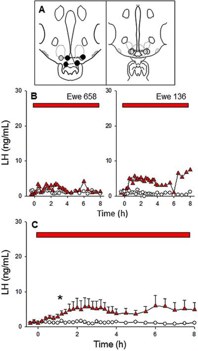
Figure 3.
Effect of microimplants of senktide in the ARC on LH secretion in follicular phase ewes. Top panel (A) depicts microimplant placement within the ARC. The middle (B) and bottom (C) panels depict LH patterns in two representative ewes and the mean (± SEM) LH concentrations for the group, respectively, during treatment with senktide-containing (red triangles) or empty (open circles) microimplants. Red bars indicate the time period of microimplant treatment. Asterisks in panel C indicate the first point at which LH concentration in senktide-treated ewes was significantly greater than in controls. Note that the scale of the y-axis is the same as Figs 1 and 2 to facilitate comparisons of the responses to senktide in all three areas.

