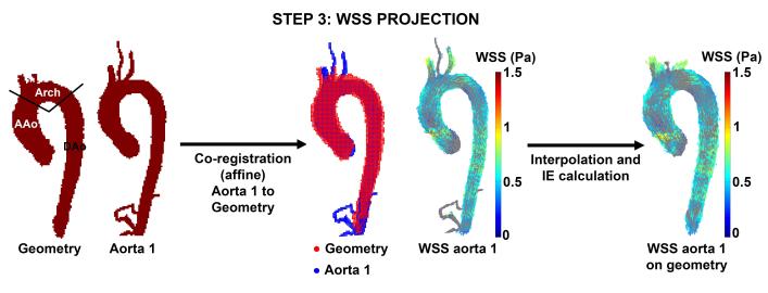
Figure 5.
WSS projection. Aorta 1 was registered to the cohort-specific aorta geometry. The WSS vectors on aorta 1 were subsequently interpolated to the aorta geometry and the interpolation error was calculated in the ascending aorta (AAo), aortic arch (Arch) and descending aorta (DAo). This step was repeated for each aorta in the cohort.

