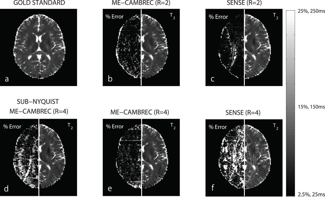
Fig 6. T2 maps from ME-CAMBREC and EPG fitting to SENSE images.
The left side of each panel is a relative difference between the given T2 map and the EPG-fit, fully-sampled, 180° refocused T2 map. Panels b, d, and e are ME-CAMBREC reconstructions, while panels c and f are T2 maps from EPG fitting to SENSE-reconstructed images. At acceleration factor 2, SENSE provides better T2 maps than ME-CAMBREC in regions with little aliasing and worse maps in regions with more severe aliasing (b,c). ME-CAMBREC scales better to higher acceleration factors than SENSE (e,f). Acceleration by the sub-Nyquist sampling of k-space combined with sub-sampling the echo dimension (d) showed a poorer result than acceleration by sub-sampling the echo dimension alone (e).

