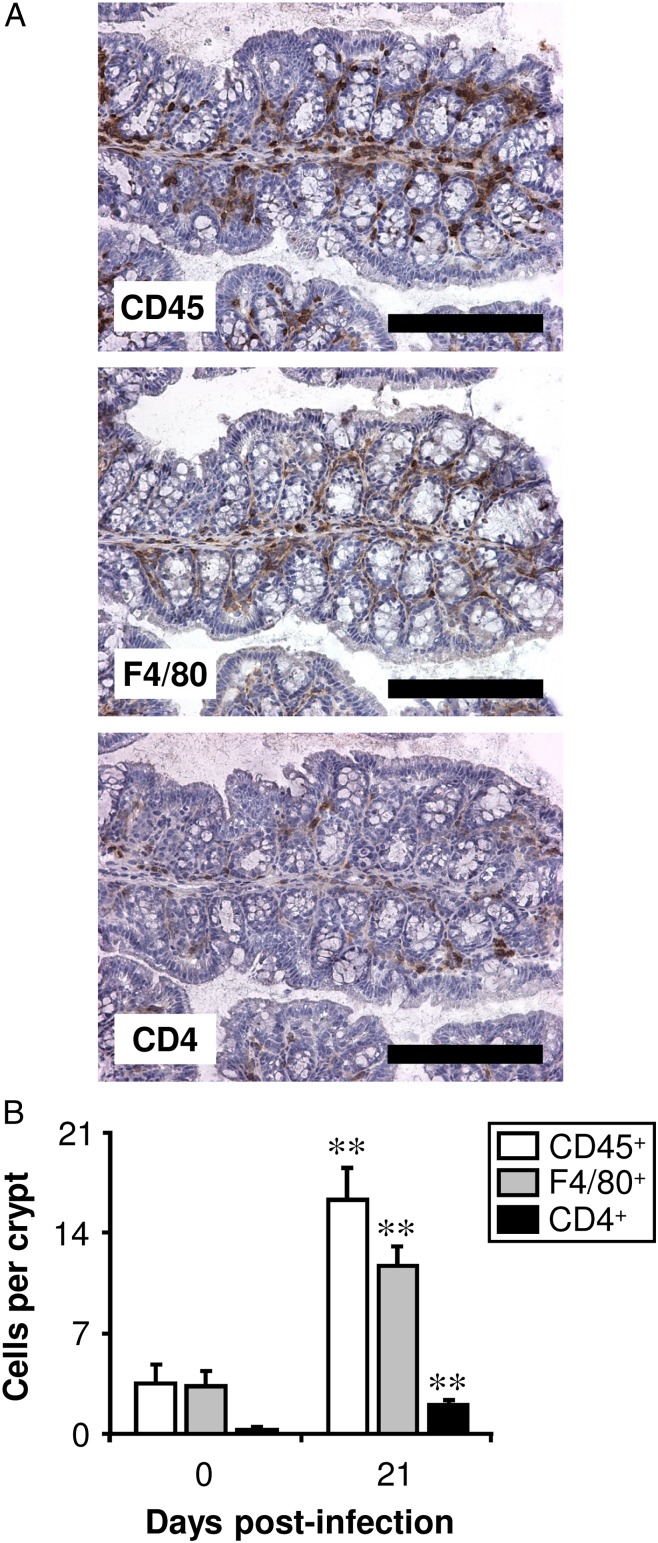
FIGURE 1.
Mφs are the predominant type of leukocyte in the large intestine both before and postinfection with T. muris. C57BL/6 mice were either left uninfected or infected with a high level of T. muris ova. Immunohistochemical staining of leukocytes (CD45+), Mφs (F4/80+), or Th cells (CD4+) was conducted on sections of the proximal colon. (A) Representative photographs are shown of serial sections from one mouse, 21 d postinfection. Scale bars, 200 μm. Quantitative analysis of the staining in both uninfected (0 d postinfection) and infected (21 d postinfection) mice is shown in (B). The values represent the means + SEM of between five and seven mice in each group, and the results are representative of three separate experiments. **p < 0.01 (21 d postinfection compared with uninfected).

