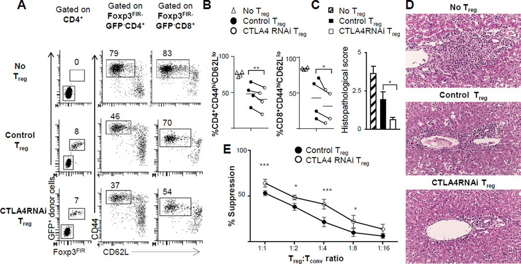
FIGURE 2. The in vivo and in vitro suppressive function of CD4+Foxp3+ Treg cells was not compromised but rather enhanced by a modest reduction in CTLA4 expression.
(A) Flow cytometry plots of the spleen of B6.Foxp3sf mice reconstituted with naïve Treg cells from CTLA4RNAi or PL4 vector control donors. Donor Treg cells were marked with ubiquitously expressed GFP in the PL4 and CTLA4RNAi transgenic lines (numbers represent percentages of gated CD4+ and CD8+ populations). (B)In vivo suppression of CD4+ and CD8+ T-cell activation determined by analyses of the CD44hiCD62Llow subset. Each data-point represents one animal (line indicating average of the group). (C–D) Histopathological scores and representative H&E sections of the livers of the B6.Foxp3sf mice reconstituted with Treg cells (original magnification: x12.5) assessed for lymphocytic infiltration (n=4 per group from 4 independent experiments; Mean ± SEM). (E) Summarized results from in vitro Treg suppression assays (n=5 from 3 independent experiments; Mean ± SEM). Control Treg cells were from transgene-negative littermates or age- & sex-matched PL4 vector transgenic mice. *p<0.05; **p<0.01; ***p<0.005.

