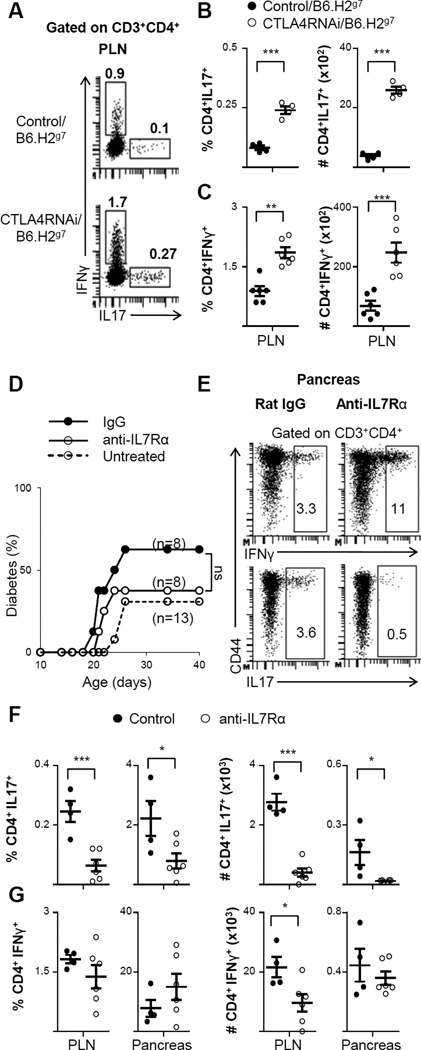
FIGURE 8. Blocking IL7 signaling with anti-IL7Rα antibody treatment suppressed Th17 formation but not diabetes development.
(A) Representative intracellular flow cytometry plots of IFNγ- and IL17-producing CD4+ T cells in the PLN (numbers represent percentages of gated populations). (B–C) Frequencies and total cell numbers of CD4+IL17+ (B) and CD4+IFNγ+ (C) subsets impacted by CTLA4RNAi on the B6.H2g7 background. Control mice were transgene-negative littermates (n=4–6 per group). (D) Littermate CTLA4RNAi/B6.H2g7 mice were treated with non-specific ratIgG2a control antibody or anti-IL7Rα antibody for 3 weeks. Mice were monitored for diabetes for 40 days. (E) Representative intracellular flow cytometry plots of the pancreas showing IL17- and IFNγ-production by CD4+ T cells after anti-IL7Rα treatment (numbers represent percentage of gated populations). (F–G) Frequencies and total cell numbers of CD4+IL17+(F) and CD4+IFNγ+ (G) after anti-IL7Rα treatment (n=4–6 mice per group). Each data-point represents one animal (Mean ± SEM). *p<0.05; **p<0.01; ***p<0.005; ns, not significant.

