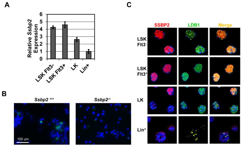
Fig 1. Abundant Ssbp2 expression and colocalization with LDB1 in murine HSPCs.
A. Ssbp2 RNA expression pattern in subpopulations of mouse BM cells as detected by real-time PCR. Cells were sorted by flow cytometry based on their surface markers, including LT-HSCs (Lin−Sca1+c-Kit+Flt3− [Flt3− LSKs]), ST-HSCs (Lin−Sca1+c-Kit+Flt3+ [Flt3+ LSKs]), progenitor cells (Lin−c-Kit+ [LKs]), and Lin+ cells. Data represent mean ± S.D. from three independent experiments.
B. SSBP2 expression is restricted to a few cells in normal BM. Mononuclear cells from whole BM were immunostained with anti-SSBP2 antibodies. Nuclear DNA was stained with DAPI (blue). Representative fields illustrate antibody specificity. A few cells in WT BM were immunoreactive. No signal was detected in null mice.
C. SSBP2 and LDB1 colocalize in the nucleus. FACS-sorted subpopulations of HSPCs were fixed, permeabilized, and stained with anti-SSBP2 and -LDB1 antibodies. Nuclear DNA was stained with DAPI.

