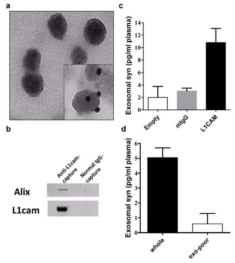
Fig. 2. Characterization of immunoaffinity-captured exosomes from human blood plasma.
(a) Electron micrograph of anti-L1CAM-captured plasma exosomes (inset: immunogold labeling of L1CAM). (b) Western blot showing that Alix, a common exosome marker, and L1CAM were enriched with anti-L1cam capture, but not with normal IgG capture. (c) α-Synuclein (syn) levels in anti-L1CAM-captured plasma exosomes were measured using a Luminex immunoassay, compared to the levels in normal mouse IgG-captured (mIgG) or “Empty” (no bead “capture”) samples. (d) Specificity was also confirmed by using exosome-poor plasma (supernatant after ultracentrifugation). Aliquots from the same pooled samples were used in these experiments (b-d) for comparison.

