
Figure 3. HS induces AMϕ autophagy via NOD2 signaling.
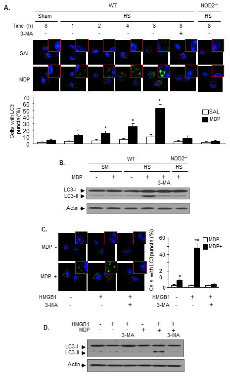
Mice were subjected to the HS-MDP two-hit model. In some animals 3-MA was injected (15 mg/kg B.W., i.t.) 10 min prior to MDP. AMϕ were then isolated from BALF at 1 - 8 h after MDP. LC3 puncta in AMϕ was detected using immunofluorescence confocal microscopy (A), and LC3-I and LC3-II in AMϕ were measured by Western blot (B). n=3/gp, mean ± SD, * P< 0.01 vs. the groups with no MDP treatment. AMϕ isolated from non-surgically treated WT mice were sequentially treated with HMGB1 for 4 h then with MDP for 8 h. In some groups, 3-MA (5 mmol/L) was added to the cells prior to MDP. LC3 puncta in the AMϕ was detected using immunofluorescence confocal microscopy (C), and LC3-I and LC3-II in AMϕ were measured by Western blot (D). Images are representative of three independent studies. n=3/gp, mean ± SD, ** (*): P<0.01 vs. indicated groups.