FIGURE 1.
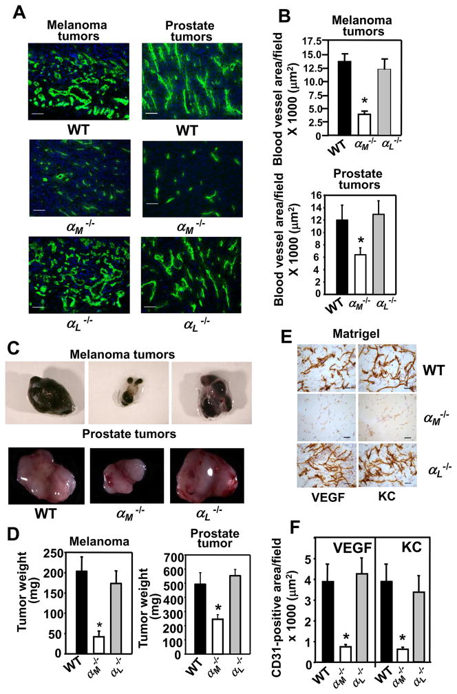
Angiogenesis is impaired in the αM−/− mice. (A) Representative images of melanoma (left panel) and prostate (right panel) tumor sections stained with an EC marker, CD31 antibody (green), Scale bars, 50 μm. (B) Decreased area of CD31-stained vasculature in tumors grown in αM−/− mice. Data are representative of two independent experiments with 8 mice per group. (C&D). Average weight of melanoma and prostate tumors grown in αM−/− is lower than in WT mice and representative tumors are shown in C. Data are means ± SEM, (n= 8 mice per group) (E) Representative images of Matrigel implant sections stained with CD31 antibody. Scale bars, 50 μm. (F) Reduced area of CD31-positive vasculature in the VEGF- or KC containing Matrigel implants from the αM−/− mice. The data are representative of 3 independent experiments with 8 mice per group.

