FIGURE 2.
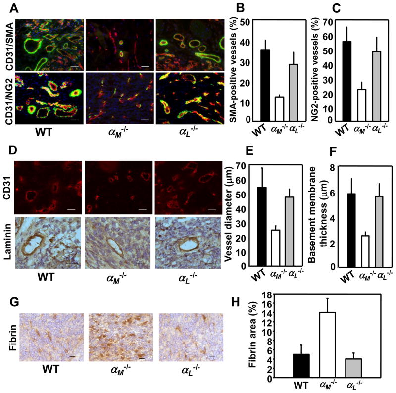
Neovasculature in prostate tumors in αM−/− mice is immature and leaky. Immunohistochemistry and image analyses of RM1 prostate tumors implanted into WT, αM−/− and αL−/− mice. (A) Costaining for SMA (green) and CD31 (red; top panel) and for NG2 (green) and CD31 (red; bottom panel) in RM1 prostate tumors. Nuclei are stained with DAPI. Scale bars, 50 μm. (B&C) Quantification of the data presented in A top and bottom panel, respectively. Data are expressed as mean ± SEM and are representative of 3 independent experiments (n=8 mice/group). (D) CD31-stained (top panel) and laminin-stained (bottom panel) blood vessels in tumors grown in WT, αM−/− and αL−/− mice. Scale bars, 50 μm (top) and 25 μm (lower panel). (E) Vessel diameter was measured in 60 tumor vessels cut perpendicular to their longitudinal axis in 8 μm-thick sections, stained for CD31 from each mouse (4 mice/group). Data are expressed as mean ± SEM, n=60. (F) Thickness of laminin-positive basement membrane in blood vessels formed in WT, αM−/− and αL−/− mice. Data are mean ± SEM, n=60 and are representative of 3 independent experiments including 4 mice per group. (G) Representative photograph of fibrin content (brown) in tumors grown in WT, αM−/− and αL−/− mice. Scale bars, 25 μm. (H) Quantification of fibrin-positive areas in tumor sections stained with anti-fibrin Ab. Data are means ± SEM, n=8 and are representative of 3 independent experiments including 8 mice per group.

