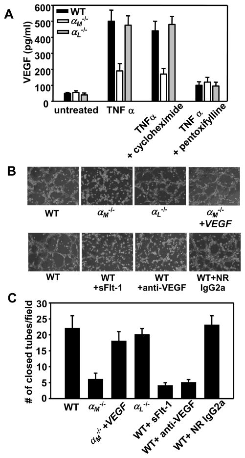
FIGURE 6.
αMβ2 supports angiogenesis via regulation of VEGF secretion by PMNs. (A) Peripheral blood WT, αM−/− or αL−/− PMNs (3×10⁶ cells/well) were incubated in 24-well TC plates in the absence or presence of TNFα (20ng/ml) for 2h at 37°C. Cycloheximide (10μg/ml) or pentoxifylline (300 μM) were added 60 min before addition of TNFα. VEGF concentration was measured in supernatants using mouse VEGF Quantikine Elisa Kit. Data are means ± SEM of triplicate samples and are representative of three independent experiments. (B) Bright field microscopy of tube formation by WT MAECs in the presence of conditioned media collected from WT, αM−/− or αL−/− peripheral blood PMNs stimulated with TNFα (upper panels). Inhibitors of VEGF: neutralizing anti-VEGF mAb, isotype matched rat IgG2a (100 μg/ml) and recombinant mouse sFLT-1 (100ng/ml) were preincubated for 60 min with conditioned media of WT TNFα-stimulated PMNs before its addition to MAECs (lower panels). The images were taken after 6h incubation in 37°C, 5% CO2. Scale bars, 75 μm. (C) Quantification of tube formation. Number of closed tubes was counted in 20 different fields of each treatment and plotted as mean ± SEM and are representative of two independent experiments.

