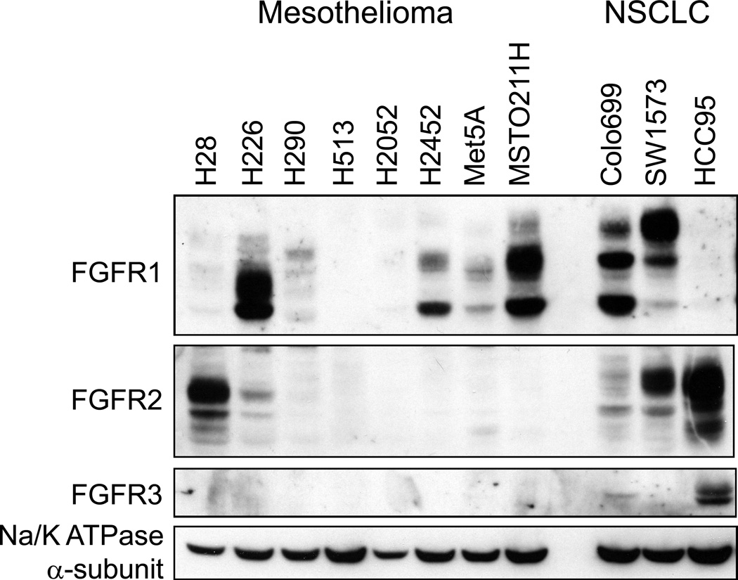
Figure 1. FGFR protein expression in mesothelioma cell lines.
Cell extracts were prepared from the indicated cell lines and aliquots were submitted to SDS-PAGE. Following electrophoretic transfer, the filters were probed for antibodies to FGFR1–3 and the α-subunit of NaK-ATPase as a loading control. Colo699 and SW1734 cells were employed as positive controls for FGFR1 and HCC95 cells are a positive control for expression of FGFR2 and FGFR3.

