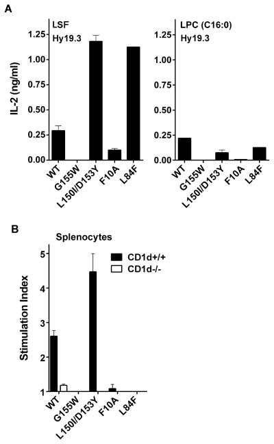
Figure 6. Modification of residues in CD1d molecules around the A′ but not F′ pocket inhibits recognition of LPC by a type II NKT cell hybridoma.
(A) In a CD1d coated plate assay Hy19.3 was stimulated in the presence of wild-type CD1d or mutant CD1d molecules with modifications in residues as indicated. IL-2 release at an optimum concentration of either 2 μg/ml of LSF or 5 μg/ml of LPC is shown. These data are representative of 3 independent experiments. (B) A proliferative response of spleen cells from naïve wild-type C57BL/6 (CD1d+/+) or CD1d-deficient (CD1d−/−) mice in response to a typical CD1d coated plate assay as above in the presence of an optimum concentration of LPC (5 μg/ml) is shown. Stimulation index was calculated by dividing CPM in the presence vs. absence of the lipid. These data are representative of 2 independent experiments.

