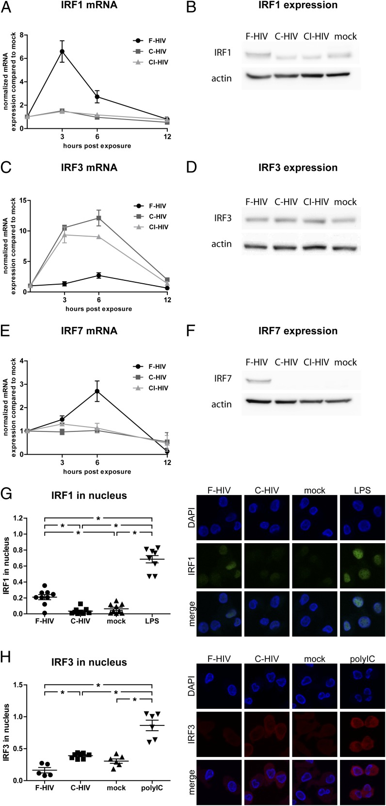
FIGURE 3.
Complement opsonization of HIV-1 suppressed IRF1 and IRF7 but activated IRF3 signaling. DCs (1 × 10⁶/ml) were exposed to F-HIV, C-HIV, and CI-HIV at an MOI of 8 or were mock treated. mRNA and protein expression of IRF1 (A and B), IRF3 (C and D), and IRF7 (E and F) was assessed using qPCR (A, C, and E) and Western blot (B, D, and F). DCs were exposed to F-HIV, C-HIV, or mock treated and the fraction of IRF1 (G) and IRF7 (H) in the cell nuclei was quantified using a confocal microscope with LPS and polyinosinic-polycytidylic acid as positive controls. Images show representative cells stained with DAPI (nuclei, blue) and IRF1 (green) or IRF3 (red) (original magnification ×40). qPCR values have been normalized with the mock value set to 1. Data are shown as means ± SEM for graphs and as representative results for confocal and Western blot images of three to six independent experiments. *p < 0.05.

