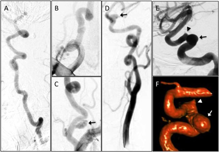
Figure 1.
Angiography demonstrating marked tortuosity and aneurysms of the vertebral (VA) and carotid arteries. Digital subtraction angiography (DSA) of the right VA (A, lateral view), right VA origin (B), left VA origin (C) and left carotid artery (D, anteroposterior view; E, lateral view; F, three-dimensional reconstruction). There is a 10 mm pseudoaneurysm along the left V1 segment (C, arrow). There is an 8 mm×10 mm wide-necked proximal cavernous left internal carotid artery (ICA) aneurysm (D–F, arrow) and a 3 mm aneurysmal irregularity of the posterior genu of the cavernous left ICA (E and F, arrowhead).

