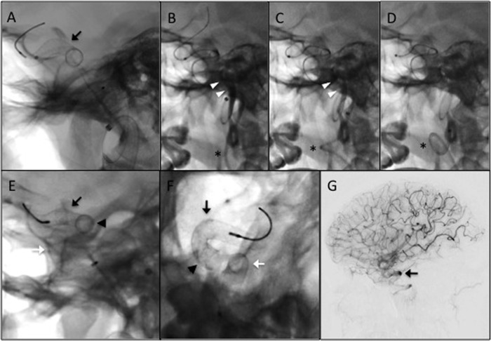
Figure 2.
Deployment of the Pipeline embolisation devices (PEDs). (A) Native fluoroscopy image demonstrating the first deployed PED (black arrow, also in E–F), sized 5 mm×18 mm. (B–D) Following deployment of the first PED, the Marskman microcatheter was readvanced over a Synchro-2 standard microwire and slack pulled from the system. These actions caused straightening of the lower cervical internal carotid artery (ICA) loop (astrisk) and contrast stasis (white arrowheads) distal to the guide catheter. The patient subsequently went asystolic, likely secondary to manipulation of the carotid bulb. The wire was quickly withdrawn and the microcatheter was reloaded, restoring the lower ICA loop and normal flow in the vessel (disappearance of contrast). Within 15 s, normal systolic cardiac rhythm restarted. Control angiography demonstrated that the first PED did not fully cover the aneurysm neck (not shown). A second PED, also sized 5 mm×18 mm, was subsequently deployed to provide full neck coverage. (E and F) Native fluoroscopy images (E, lateral; F, anteroposterior) following deployment of the second PED (white arrow). The arrowhead in (E) and (F) represents the region of overlap between the two telescoping PEDs. (G) Control digital subtraction angiography immediately postdeployment of the second PED demonstrating contrast stasis in the aneurysm (black arrow).

